
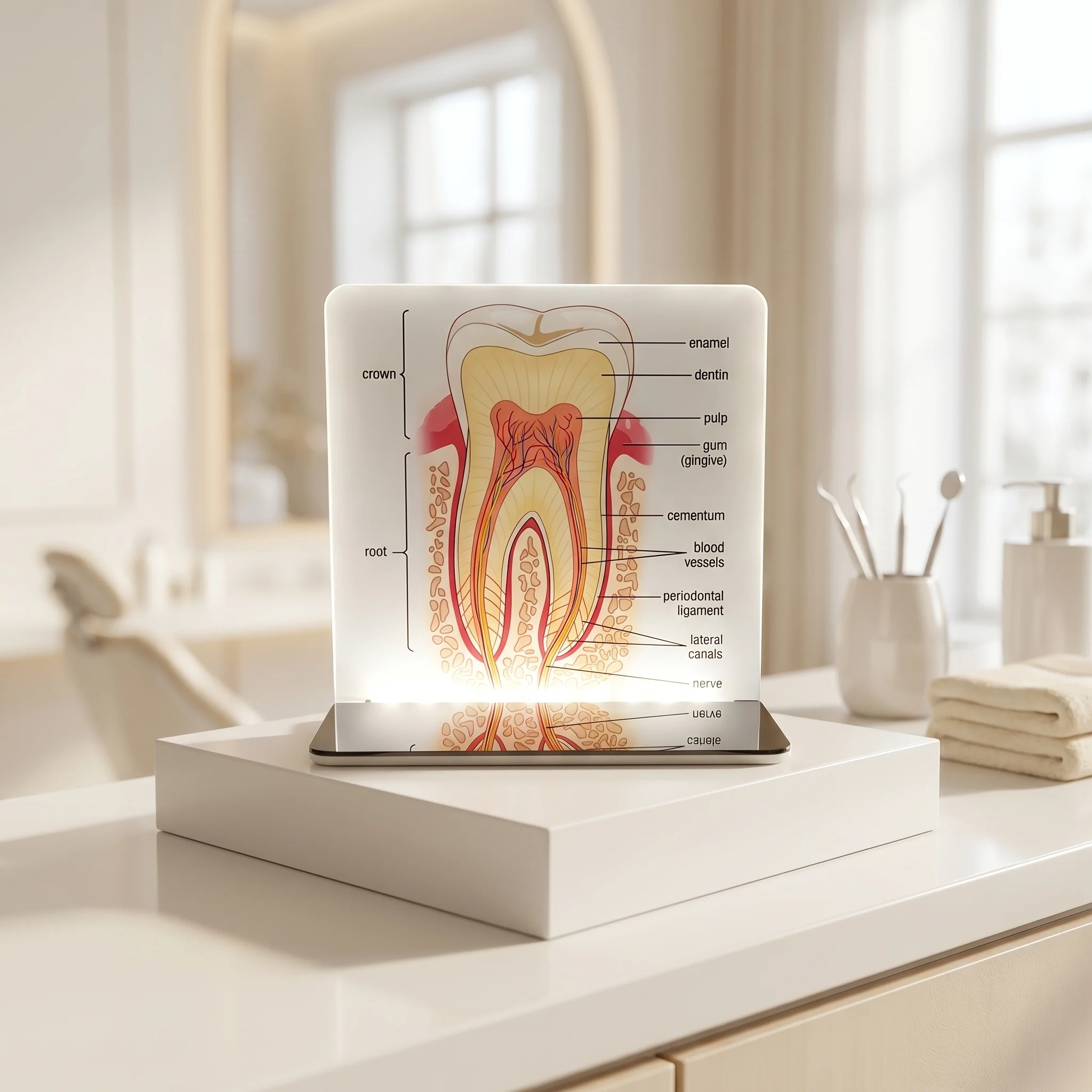
Dental Desk Anatomy Model
Original price was: ₨ 2,550.00.₨ 1,947.00Current price is: ₨ 1,947.00.
Enhance your clinic’s workspace with the Dental Desk Anatomy Model, available exclusively at Dental Needs.Pk
- Sleek & Space-Saving: Modern flat-panel design that fits perfectly on any countertop.
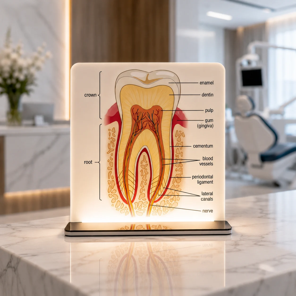
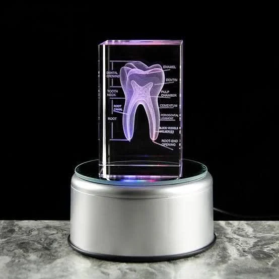
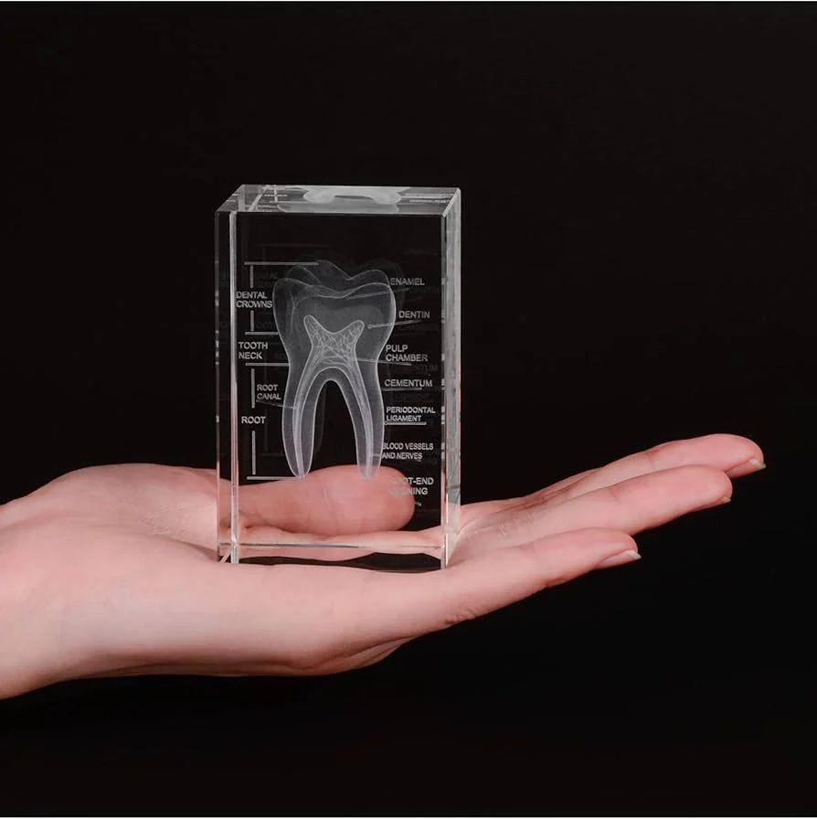
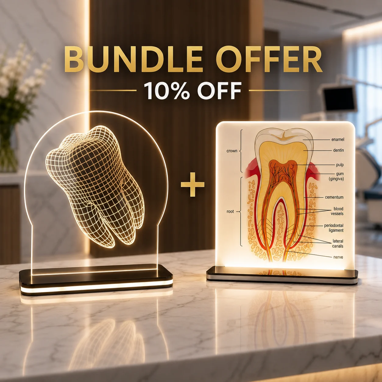
- Patient Education Tool: Clearly labels enamel, dentin, pulp, and root structure to help explain procedures easily.
- Premium Illumination: The LED base casts a brilliant, professional glow that elevates your clinic’s aesthetic.
Upgrade your desk setup—order yours today from DentalNeeds.Pk!

Estimate delivery times: 3-5 days all over Pakistan.

Free Shipping: On Online Payment.
Dental Desk Anatomy Model – Illuminated Acrylic Patient Education Display
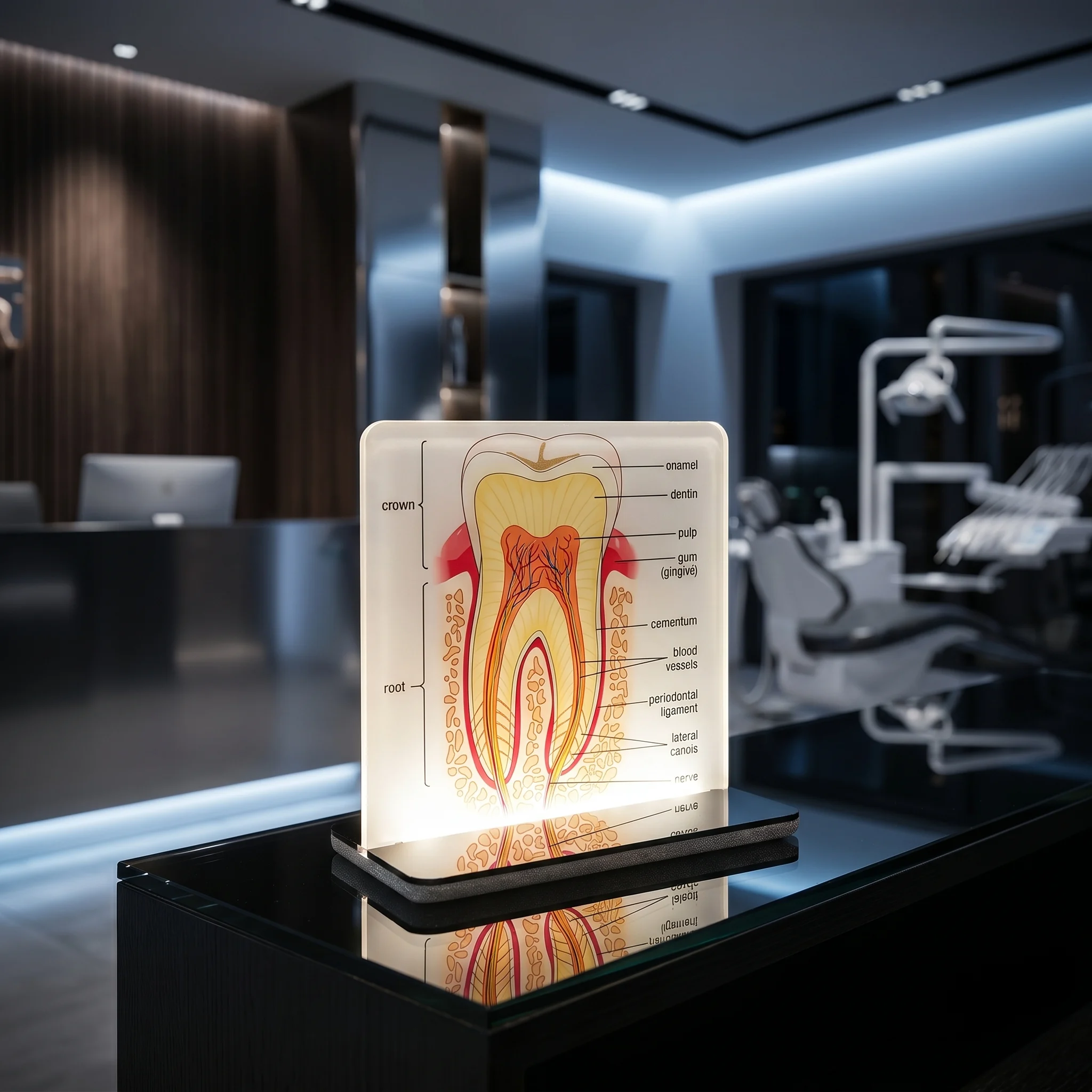
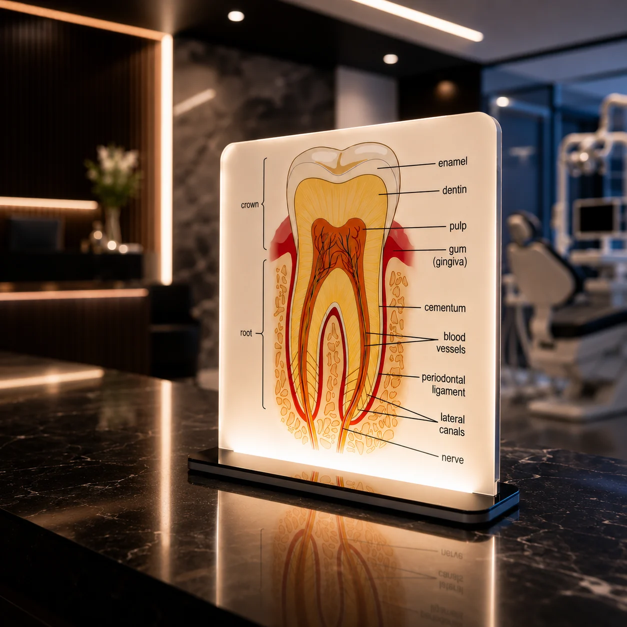
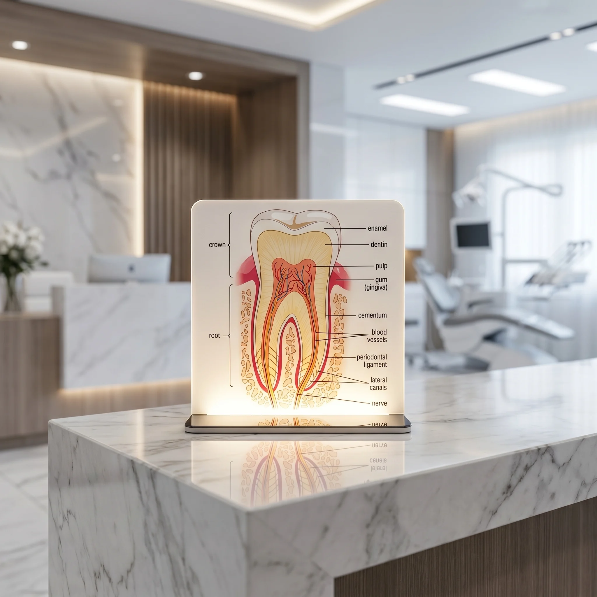
Enhance your clinic’s reception or consultation desk with the Dental Desk Anatomy Model, exclusively available at Dental Needs.Pk This sleek, modern, flat-panel acrylic display features a highly detailed, colorful cross-section of a tooth, beautifully illuminated by a premium LED light base.
Key Features
-
Crystal-Clear Acrylic Panel: Made from high-grade, durable flat acrylic that beautifully diffuses light while maintaining a clean, glass-like premium look.
-
Vibrant Tooth Cross-Section: Features accurate anatomical labeling—including the enamel, dentin, pulp, gum (gingiva), cementum, blood vessels, periodontal ligament, lateral canals, and nerve—making it the perfect visual aid.
-
Premium Illuminated Base: The sleek bottom-lit LED base casts a brilliant, professional glow upwards through the panel, catching the eye of every patient.
-
Compact Countertop Design: Fits perfectly on any dental reception desk, consultation table, or office shelf without cluttering the workspace.
Why Your Clinic Needs This Model
-
Simplify Patient Communication: Don’t just explain root canals, cavities, or crown procedures—show them. This clear visual guide helps build trust and patient compliance during treatment planning.
-
Elevate Clinic Aesthetics: Move away from bulky, old-school plastic molds. This modern, flat-panel glowing display gives your clinic a tech-forward, premium vibe.
-
The Perfect Gift for Dentists: An excellent, sophisticated gift for graduating dental students, colleagues, or opening ceremonies of new dental practices.
🦷 Detailed Anatomical Markings Included:
The Dental Desk Anatomy Model features a complete, highly accurate cross-section breakdown divided into two main sections:
-
The Crown: Highlights the upper visible portion of the tooth.
-
Enamel: The hard, protective outer layer.
-
Dentin: The supportive sensitive layer beneath the enamel.
-
Pulp: The central chamber housing nerves and blood vessels.
-
Gum (Gingiva): The surrounding soft tissue.
-
-
The Root: Showcases the internal structure beneath the gum line.
-
Cementum: The protective layer covering the tooth root.
-
Blood Vessels & Nerves: Clearly mapped out to demonstrate tooth vitality.
-
Periodontal Ligament: The connective tissue fibers attaching the tooth to the bone.
-
Lateral Canals: Demonstrating complex root canal anatomy.
-
| Weight | 0.5 lbs |
|---|
Add a review
Related products
Clinic Decor
Clinic Decor
Clinic Decor
Clinic Decor
Clinic Decor
Dental Models
6 reviews for Dental Desk Anatomy Model